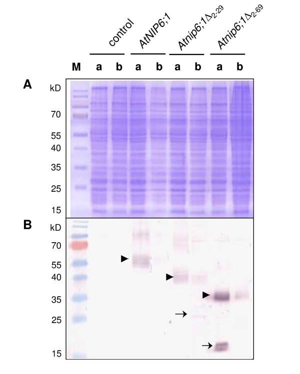
Figure 6.
Detection by Western blotting of AtNIP6;1, Atnip6;1Δ2–29 and Atnip6;1Δ2–69 expressed in yeast. We ran 20 μg of microsomal membrane proteins from (a) Δfps1 and (b) Δfps1 Δacr3 Δycf1 transformed with the empty vector or AtNIP6;1, Atnip6;1Δ2–29 or Atnip6;1Δ2–69 on 12% SDS polyacrylamid gels. Proteins were (A) stained with commassie brilliant blue or (B) transferred to nitrocellulose membrane for probing with an anti-AtNIP6;1 specific antibody. M, PageRuler™ Prestained Protein Ladder (Fermentas). The arrows point towards bands that probably represent monomeric forms of Atnip6;1Δ2–29 and Atnip6;1Δ2–69. Arrowheads point towards bands that probably represent oligomeric forms of the proteins.

