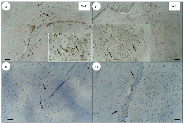
Figure 4.
Immunochemical staining of the glial fibrillary acidic protein (GFAP) in mouse brain. Astrogliosis with apparent GFAP expression (arrow) was observed in the parenchyma near to the choroid plexus in the brains of infected mice at 4 wpi (A) or 8 wpi (C). However, weak GFAP expression was also detected in the brains of age-matched uninfected mice at 4 wpi (B) and 8 wpi (D). Bar = 50 μm. Inserts are higher magnifications of GFAP expression from the same panel.

