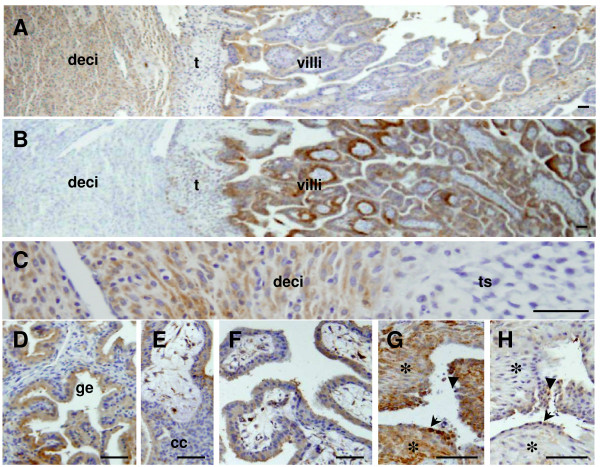
Figure 7.
HTRA3 immunolocalisation at the maternal-fetal interface. A, a low power view of an implantation site. C, a higher power view of A showing the decidual-trophoblast shell junction. D, maternal glands. E, anchoring villi, F, floating villi. Cytokeratin staining in implantation sites is shown in panels B and G. B, an adjacent section of A stained for cytokeratin; G, extravillous endovascular trohoblasts (EVTs) surrounding a blood vessel stained for cytokeratin; EVTs outside the vessel are highlighted by asterisks (*), and those lining and inside the vessel by the arrow and arrowhead respectively. H, an adjacent section of G stained for HTRA3. Deci, decidual cells; t, trophoblast cells; ts, trophoblast shell; ge, glandular epithelium; cc, trophoblast cell column. Scale bar = 50 μm.

