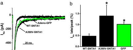
Fig. 5.
A390V-SNTA1 increased late INa in native cardiac cells compared with that of WT-SNTA1. (a) Whole-cell INa traces were elicited by step depolarization of 775 ms in duration to −20 mV from a holding potential of −140 mV and normalized to cell capacitance. Adenoviral recombinants of WT-SNTA1, A390V-SNTA1-Ires-GFP, and GFP alone were used to transduce neonatal cardiomyocytes for 48 h. (b) Summary data for the percentage of late INa at 775 ms normalized to peak current.

