
Figure 3. No Significant Neuron Loss Was Associated with n-3 Polyunsaturated Fatty Acid Depletion in the Tg2576 Mouse.
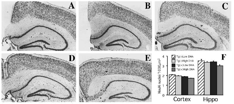
(A–E) Immunostaining with NeuN, an antibody that labels neuronal nuclei, revealed similar neuronal counts in the hippocampus or cortex between Tg2576 mice fed with low n-3 PFA diet [Tg(+) Low DHA] (A), Tg(−) fed with low n-3 PFA diet [Tg(−) Low DHA] (B), Tg(+) and Tg(−) mice fed with DHA-enriched diets (High DHA) ([C] and [D], respectively), and Tg2576 mice raised on control chow (E). Original magnification, 100×. (F) Image analysis quantification of NeuN-ir (number of neuronal nuclei per 1000 μm2) was performed on neuronal layers in cortex (entorhinal II, entorhinal III/IV, parietal II, parietal III/IV, parietal V/VI, frontal II, frontal III/IV, frontal V/VI) and hippocampus (CA1, CA2, CA3) at Bregmas −1.0 mm, 1.7mm, −2.7 mm, −3 mm (analyzed from anterior to posterior hippocampus with four consecutive sections analyzed per Bregma). There were no significant neuron density changes with APPswe transgene or with diet (n = 5 or 6 in each group). Since there were no treatment effects on neuronal nuclei densities in any layer and no interaction with regions, we show interaction bars rather than the breakdown of densities in layers and different Bregmas.