Figure 6.
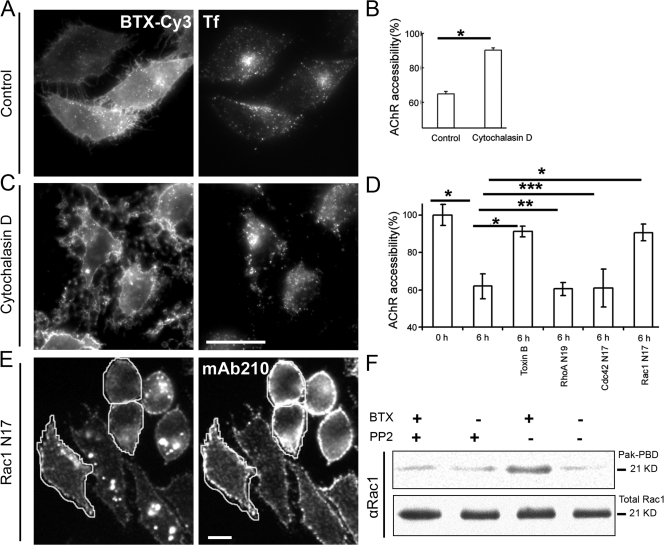
Inhibition of actin dynamics or Rho GTPase Rac1 blocks AChR sequestration and endocytosis. (A–C) CHO-K1/A5 cells were labeled with b-Cy3αBTX in the absence (control) or presence of cytochalasin D for 2 h and were labeled at 4°C with Cy5-SA to assess the fraction of surface-accessible receptors as described in Materials and methods. Comparison of Tf-AlexaFluor488 uptake (right panels in A and C) in control and cytochalasin D–treated cells shows that cytochalasin D treatment did not affect Tf-AlexaFluor488 uptake. The histogram in B shows the extent of Cy5-SA accessibility in cytochalasin D–treated and control cells normalized to that obtained at 0 h. Note that surface accessibility of AChR increased significantly in cytochalasin D–treated cells compared with control cells exposed to DMSO alone. Mean and SD (error bars) were obtained from at least 60 cells per experiment; similar results were obtained in three independent experiments. *, P < 0.001. (D) Cells were transfected with different dominant-negative mutant isoforms of Rho GTPases (Cdc42 N17, RhoA N19, and Rac1 N17) for 14 h or were treated for 1 h with 1 μg/ml C. difficle toxin B, were labeled with αBTX, subsequently chased for 6 h at 37°C, and incubated with mab 210 followed with secondary antibody on ice for 1 h to determine the level of surface AChR remaining at the end of the chase period. The histogram shows weighted mean ± SEM (error bars) values from two independent experiments. *, P < 0.0001; **, P > 0.05; ***, P > 0.1. (E) Images show the distribution of internalized b-Cy3αBTX (left) and mAb 210 antibody–stained surface AChR (right) in cells transfected with Rac1 N17 (outlined) with respect to untransfected control cells. (F) αBTX incubation of AChR-expressing CHO-K1/A5 cells with or without 10 μM PP2 were lysed, and activated Rac1 was precipitated using PAK-PBD; total Rac1 and PAK-PBD–precipitated Rac1 were analyzed by Western blots from the same cell lysates. Note that αBTX binding induces PP2-inhibitable Rac1 activation. Bars: (C) 20 μm; (E) 10 μm.

