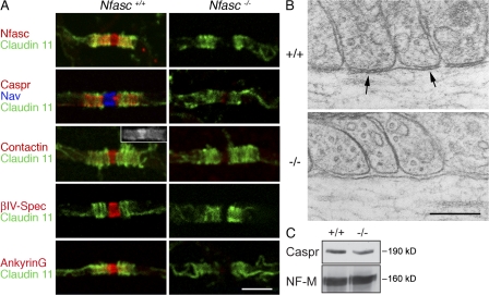
Figure 1.
Disruption of CNS paranodes and nodes in Nfasc−/− mice. (A) Immunofluorescence analysis of teased fibers from the ventral funiculus of the cervical spinal cord from Nfasc+/+ and Nfasc−/− animals showed that nodes and paranodes were disrupted when Nfasc155 and Nfasc186 were lost. Immunostaining of teased fibers for Claudin 11 (Oligodendrocyte-specific protein) was used to localize the paranodes of CNS myelin. Immunostaining with an antibody that recognized both Nfasc186 and Nfasc155 (Nfasc) confirmed that they were no longer at the node and paranode, respectively. The axoglial junctional components Caspr and Contactin were also no longer concentrated at the paranodes. In the CNS, Contactin is present at both the node and paranodes (inset). The nodal components voltage-gated sodium channels (Nav), Contactin, βIV-Spectrin (βIV-Spec), and AnkyrinG were mislocalized in mutant animals. Note that AnkyrinG in the CNS has both a nodal and paranodal localization. Bar, 5 μm. (B) Electron microscopy of the paranodes in wild-type (+/+) and mutant (−/−) mice. Septate junctions at the base of paranodal loops (arrows) were no longer present in the mutant. Bar, 0.2 μm. (C) Western blot analysis of cervical cord homogenates from wild-type (+/+) and Nfasc−/− (−/−) mice, respectively, showing that absence of the Neurofascins does not affect the amount of Caspr in the tissue. Neurofilament-M (NF-M) was used as a loading control.

