Figure 1.
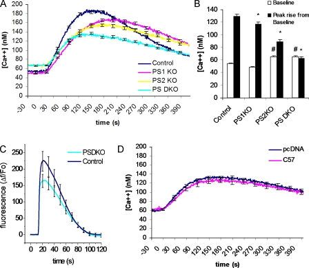
Elevated cytosolic Ca2+ and reduced ER Ca2+ stores in presenilin-null cell line. (A) Changes in cytosolic Ca2+ evoked by thapsigargin in control fibroblasts (control; n = 48 cells) and fibroblasts from PSDKO (n = 57 cells), PS1KO (n = 48), and PS2KO (n = 56) mice. ER Ca2+ stores were released into the cytosol by application of 1 μM thapsigargin, a specific blocker of SERCA activity, with 0 mM Ca2+ in the bathing solution. Basal cytosolic Ca2+ levels were elevated (∼70 nM) in the PSDKO and PS2KO fibroblasts compared with controls and PS1KO cells (∼50 nM; P < 0.05), whereas the peak Ca2+ signals after application of thapsigargin were substantially reduced in PSDKO and PS2KO fibroblasts. (B) Mean values of basal cytosolic [Ca2+] and thapsigargin-evoked Ca2+ signals, derived from the experiments in A. *, significance in peak rise versus pcDNA (P < 0.05); #, significance in basal levels versus pcDNA (P < 0.05). (C) Changes in cytosolic Ca2+ evoked by 1 μM ionomycin in control fibroblasts (control; n = 12 cells) and fibroblasts from PSDKO mice (n = 37 cells), with 0 mM Ca2+ in the bathing solution. (D) Cytosolic Ca2+ signals in PSDKO fibroblasts transfected either with pcDNA (n = 63 cells) or AICD (n = 54 cells) 48 h earlier. No significant differences were apparent in either basal cytosolic Ca2+ levels or the peak response after application of 1 μM thapsigargin. Error bars show SEM.

