Figure 1.
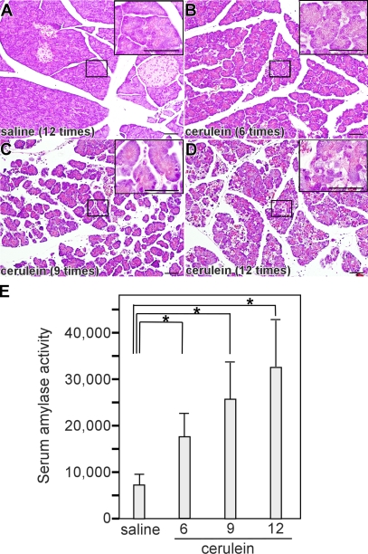
Autophagy induction in pancreatic acinar cells of cerulein-induced acute pancreatitis. Overnight-starved mice were treated by saline (A) or cerulein 6 (B), 9 (C), or 12 (D) times, and pancreatic sections were analyzed by H&E staining. Insets show higher magnifications of areas indicated in A–D. (E) Serum amylase activity in mice with cerulein-induced pancreatitis. Data are shown as mean ± SEM (error bars). *, P < 0.05. Bars, 50 μm.

