Figure 2.
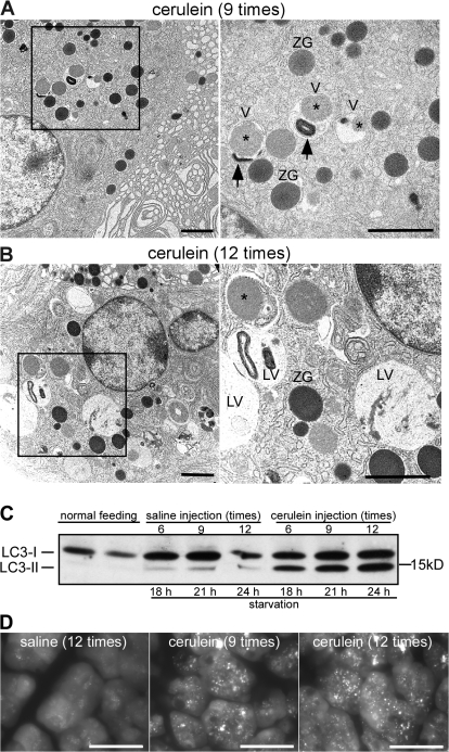
Appearance of autophagic vacuoles in cerulein-induced pancreatitis. (A and B) Mice were injected with cerulein 9 (A) or 12 (B) times, and the pancreas was analyzed by EM. The right panels show higher magnification of the boxed areas. V, autophagic vacuole; ZG, zymogen granule; asterisk, zymogen granule contained in vacuole; arrow, membrane-bound organelle contained in vacuole; LV, large vacuole. (C) LC3 conversion in acute pancreatitis. Pancreas homogenates were prepared from cerulein-injected mice and subjected to immunoblotting using anti-LC3 antibody. The cytosolic LC3-I protein (16 kD) was converted into LC3-II (14 kD), and the amount of LC3-II was correlated with the extent of autophagosome formation. (D) GFP-LC3 mice were treated with cerulein and analyzed by fluorescence microscopy. Bars: (A and B) 2 μm; (D) 10 μm.

