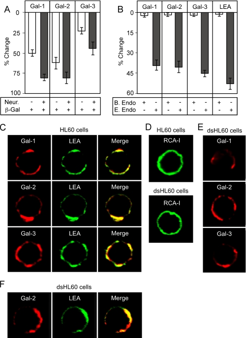
FIGURE 9.
Gal-1, Gal-2, and Gal-3 recognize poly(LacNAc) glycans on HL60 cells. A, Gal-1, Gal-2, and Gal-3 binding toward HL60 cells following treatment with jack bean β-galactosidase with or without pretreatment of cells with A. ure-afaciens neuraminidase. B, Gal-1, Gal-2, and Gal-3 binding toward HL60 cells following treatment with either B. fragilis or E. freundii endo-β-galactosidase. Bars represent the percent change in cell surface binding when compared with the mean fluorescent intensity of nontreated cells ± S.D. C, confocal analysis of Gal-1, Gal-2, Gal-3, and LEA binding toward cell surface glycans on HL60 cells. D, confocal analysis of RCA-I binding toward cell surface glycans on HL60 cells or HL60 cells treated with 100 milliunits A.ureafaciens neuraminidase (dsHL60). E, confocal analysis of Gal-1, Gal-2, Gal-3 binding toward cell surface glycans on HL60 cells treated with 100 milliunits of A. ureafaciens neuraminidase (dsHL60). F, confocal analysis of Gal-2 and LEA binding toward cell surface glycans on HL60 cells treated with 100 milliunits of A. ureafaciens neuraminidase (dsHL60).

