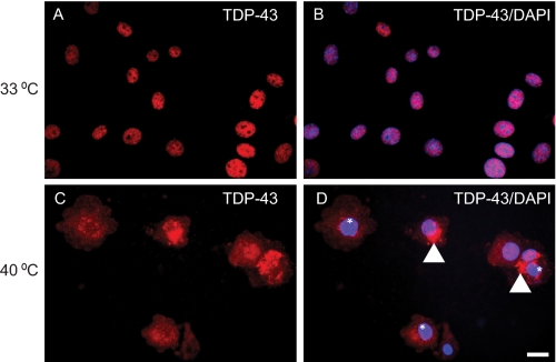
FIGURE 2.
TDP-43 expression and accumulation in cytoplasm of temperature-sensitive BN2 cells. A and B, endogenous TDP-43 (red) was detected in the nucleus of tsBN2 cells at 33 °C (permissive temperature). The merge image (B) of TDP-43 (red) and nuclei stain DAPI (blue) confirms that TDP-43 is in the cell nucleus. C and D, at nonpermissive temperature (39.5 °C), TDP-43 (red) was detected as punctate cytoplasmic aggregates (arrowheads) with clearing of nuclear TDP-43 (*) and did not colocalize with DAPI (blue) in tsBN2 cells. D, merged image. Scale bars, 20 μm.

