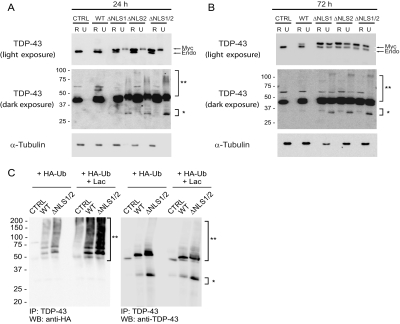
FIGURE 6.
Expression of Myc-TDP-43-ΔNLS mutants results in the sequestration of ubiquitinated and insoluble endogenous TDP-43. QBI-293 cells 24 h (A), or 72 h (B) post-transfection with empty vector (CTRL), Myc-TDP-43-WT (WT), Myc-TDP-43-ΔNLS1 (ΔNLS1), Myc-TDP-43-ΔNLS2 (ΔNLS2), or Myc-TDP-43-ΔNLS1/2 (ΔNLS1/2) sequentially extracted with RIPA (R) and urea buffer (U). Immunoblotting was conducted with TDP-43 antibody. Myc-TDP-43 (Myc) migrates slower than endogenous TDP-43 (Endo). Over-exposure of the immunoblot demonstrates the presence of a high Mr smear (**) and C-terminal fragments (*) in the urea fractions of Myc-TDP-43-NLS mutants in transfected cells. α-Tubulin was used as a loading control. C, immunoblots (IB) of immunoprecipitated (IP) QBI-293 cell lysates cotransfected with empty vector, Myc-TDP-43-WT, or Myc-TDP-43-ΔNLS1/2 and HA-tagged ubiquitin (HA-Ub) in the presence (+LAC) or absence of LAC. Note the presence of the LAC-dependent, ubiquitinated TDP-43 positive high-Mr smear and the TDP-43 C-terminal fragments.

