FIGURE 7.
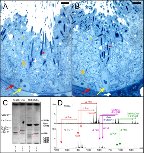
Histological (A and B) and sphingolipid (C and D) analysis of fertile Slc35c1-/- and corresponding control (Slc35c1+/+) testes. A and B, no significant changes were observed in the architecture of the seminiferous epithelium of mutant mice testes (B) as compared with controls (A). Note, maturating spermatozoa (red asterisks) are present in both control and mutant testis. Semithin Epon sections were stained with methylene blue-Azur II. White arrows, Leydig cells; yellow arrows, Sertoli cell nuclei; red arrows, spermatogonia; short orange arrows, adluminal primary pachytene spermatocytes; orange asterisks, round spermatids; bar, 10 μm. C, TLC of testicular GSLs from control (Slc35c1+/+) and Slc35c1-/- testis. GSLs were split into a neutral and an acidic fraction and stained after separation with orcinol. Lanes correspond to 20 mg of tissue wet weight. Red lanes denote the shift in migration observed for GSLs of mutant mice. Instead of bands for FucGA1 and FucGM1, bands for GA1(Gg4Cer) GM1 are observed in mutant mice. D, characterization of neutral complex GSLs from control (Slc35c1+/+) and Slc35c1-/- testes by nano-electrospray ionization-tandem mass spectrometry. Complex neutral GSLs were detected with a precursor ion scan of m/z +204. Signals for fucosylated VLCPUFA-GSLs (FucGA1, Gal(Fuc)GA1, GalNAc- (Fuc)GA1, and GalNAcGal(Fuc)GA1) dominate in control sample. In mutant (Slc35c1-/-) testis, signals for fucosylated VLCPUFA-GSLs are absent. Instead, corresponding signals for nonfucosylated GSLs appear, as measured by a down shift of 146 atomic mass units.

