FIGURE 4.
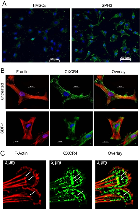
Intracellular localization of CXCR4. Cells were dissociated by trypsinization, plated on Lab-Tek II chamber CC2 glass slides, and allowed to attach for 8 h. After serum starvation, cells were treated with and without 1 μg/ml SDF-1α for 45 min. hMSCs from a monolayer (hMSCs) or 3-day-old hMSC spheroids (SPH3) were stained with anti-human CXCR4 antibody and Alexa Fluor 488-conjugated F(ab′)2 secondary antibody (green). F-actin was stained with Alexa Fluor 594-conjugated phalloidin (red). The nuclei were counterstained with 4′,6-diamidino-2-phenylindole (blue). A shows CXCR4 staining (green) in hMSCs and SPH3. B shows CXCR4 (green) and F-actin (red) staining in SPH3 cells before (untreated) and after treatment with 1 μg/ml SDF-1α for 45 min (SDF-1). C shows staining for CXCR4 (green) and F-actin (red) in lamellipodias of cells from SPH3 treated with SDF-1. Sites of co-localization are denoted by white arrows.

