FIGURE 5.
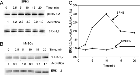
Effects of SDF-1 on activation of ERK-1,2. Cells from a monolayer of hMSCs (hMSCs) or 3-day-old hMSC spheroids (SPH3) were dissociated, plated, and allowed to adhere for 8 h. After serum starvation, cells were treated with 1 μg/ml SDF-1α for 0–20 min and lysed. Proteins were separated on an SDS-PAG gel and analyzed by Western blot using antibodies against total ERK-1,2 and pERK-1,2. A shows pERK-1,2 and total ERK-1,2 staining for cells isolated from SPH3. B shows pERK-1,2 and total ERK-1,2 for hMSCs. C shows the results of densitometric analysis of pERK-1,2 for hMSCs and SPH3 after normalization for total ERK-1,2 in corresponding cellular lysates.

