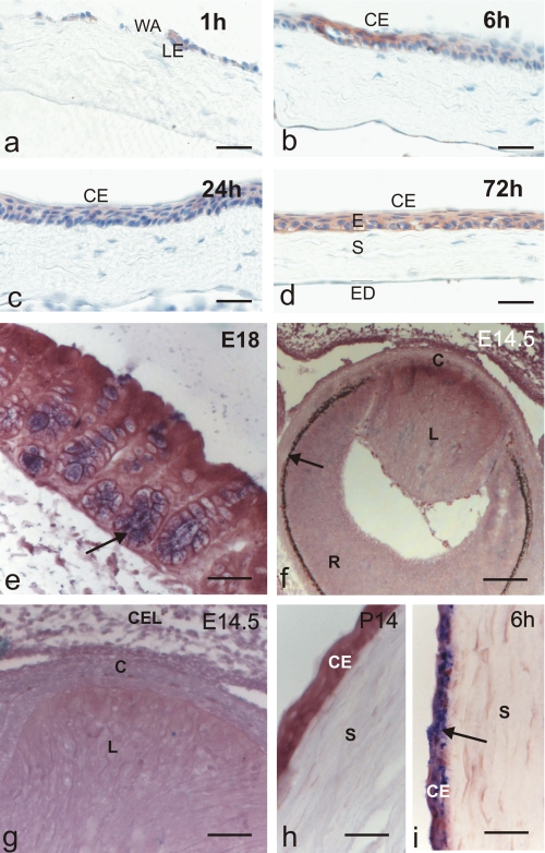
FIGURE 1.
Tff3 protein is expressed in injured corneal epithelium, but Tff3 mRNA is absent during corneal development. Immunohistochemical analysis of Tff3 peptide expression in the course of corneal wound healing after alkali burn (a-d). Wounded corneas were allowed to partially heal in vivo and were then subjected to immunostaining. Tff3 immunoreactivity was detected at the leading edge (LE) of migrating epithelium after 1 h and covering corneal epithelium (CE) up to 72 h after injury. WA, wounded area; E, epithelium, S, stroma; ED, endothelium. In situ hybridization revealed that Tff3 mRNA is expressed during development of wild type mice in goblet cells of colon mucosa (e, arrow, blue) but is lacking during corneal development (f-h). f demonstrates a section through the developing eye at E14.5. The arrow marks retinal pigment epithelium, R, developing retina; L, developing lens; C, developing cornea. g is a magnification of f indicating that no Tff3 transcripts are visible in the developing cornea (C), which is bordered by the developing lens (L) and closed eye lids (CEL) at this stage of development. h shows corneal epithelium (CE) and corneal stroma (S) at P14 lacking Tff3 transcripts. i, Tff3 mRNA is expressed in injured corneal epithelium (arrow, blue)inthe course of corneal wound healing after alkali burn. Wounded corneas were allowed to partially heal in vivo for 6 h and were then subjected to in situ hybridization. Scale bars:48 μmin a-d;32 μmin e; 82.5 μmin f;32 μmin g-i.

