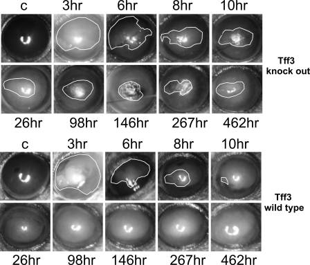
FIGURE 7.
Re-epithelialization of corneal wounds in Tff3-/- mice is prolonged in in vivo model. Representative images showing the corneal wound areas (surrounded by white lines) of wild type (upper panel) and Tff3 null mice (bottom panel) taken during the healing period under in vivo conditions at different time points. The first picture in each panel illustrates an unwounded cornea (C, control) of the contralateral eye. In wild type cornea the epithelial wound closure is completed by 10 h after injury, whereas in Tff3 knock out mice wound closure is protracted up to 462 h.

