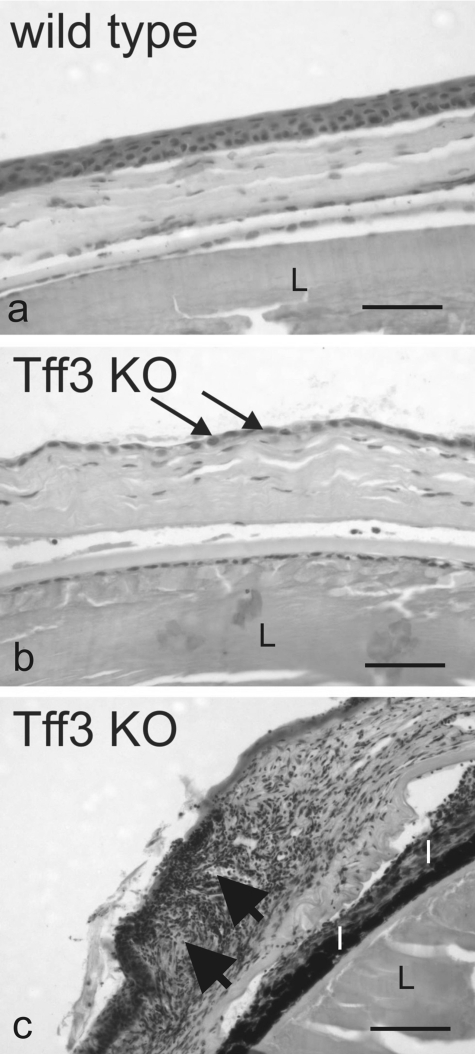
FIGURE 8.
Defective corneal epithelial wound healing in Tff3-deficient mice. Shown are light micrographs taken from the cornea of wild type (a) and Tff3 null (-/-) mice (Tff3 KO, b and c) after macroscopic epithelial wound closure. Histological analysis by hematoxylin and eosin staining of wild type and Tff3-/- mice cornea demonstrates that in Tff3-/- cornea the multilayered organization of the epithelium is thinned to a single layer of epithelial cells (b, arrows) covering the wounded area. Big arrows point to an area of the Tff3-/- cornea infiltrated by immune cells (c). I, iris; L, lens. The scale bar equals to 184 μm.

