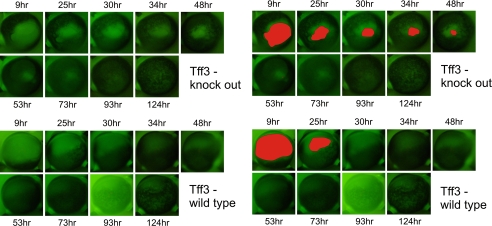
FIGURE 9.
Re-epithelialization of corneal wounds in Tff3 null mice is prolonged in combined in vivo (12 h)/in vitro model. After alkali-induced corneal wounding and healing in vivo for 12 h, the epithelial defects were stained with fluorescein. Representative images from the cornea of wild type (upper panel) and Tff3 knock out mice (bottom panel) at several time points during wound healing are shown. In the left panels the corresponding remaining wound areas are filled with red color. The epithelial wound in the Tff3 null cornea remains open by 50 h, whereas in wild type cornea the wound is completely closed by 30 h.

