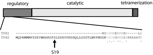
FIGURE 1.
Schematic diagram of amino acid hydroxylases. Shown is a diagram of the major domains of amino acid hydroxylases and a sequence alignment (ClustalW) of the first 60 amino acids of human TPH2 with the corresponding residues of human TPH1. Identity is indicated with an asterisk, conserved substitutions are indicated with a colon, and semiconserved substitutions are shown with a period. The candidate PKA site, Ser19, is indicated.

