FIGURE 3.
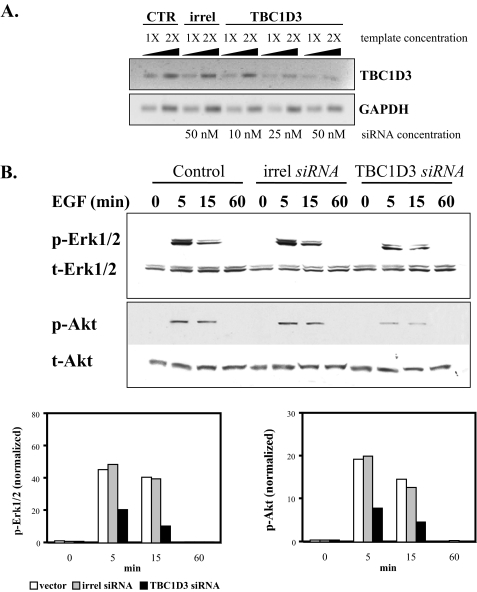
TBC1D3 depletion decreases Akt and Erk1/2 activation. A, semi-quantitative RT-PCR. TBC1D3 was suppressed in DU145 cells using a commercial siRNA pool (Dharmacon) as described under “Experimental Procedures.” Transcript levels of TBC1D3 (top panel) were determined by semi-quantitative RT-PCR from control cells (lanes 1 and 2), cells transfected with irrelevant siRNA (lanes 3 and 4) and cells transfected with TBC1D3 siRNA (lanes 5-10). B, DU145 cells were transfected with TBC1D3 siRNA (50 nm), irrelevant siRNA (irrel) or untransfected (Ctr). 36 h later the cells were serum-starved, incubated with EGF (100 ng/ml) at 37 °C for different time points, and analyzed by Western blot. The bar graphs were derived from densitometric analysis and show p-Erk1/2 and p-Akt normalized to the actual protein loaded.

