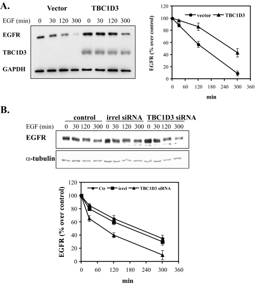
FIGURE 5.
EGFR degradation is delayed in cells expressing TBC1D3. A, DU145 cells transfected with pCMV-Myc-TBC1D3 or vector alone were serum-starved and incubated with 100 ng/ml EGF at 37 °C for the indicated times. The cells were washed, and lysates (10 μg of protein each) were analyzed by Western blotting. B, TBC1D3 depletion accelerates EGFR degradation. DU145 cells were transfected with a commercial siRNA pool (Dharmacon) (50 nm), irrelevant siRNA (irrel) or untransfected (Ctr). 36 h later the cells were serum-starved and incubated with 100 ng/ml EGF at 37 °C for the indicated times. The cells were lysed and analyzed by Western blotting. Data are presented as a ratio of the amount of EGFR present at each time point over the amount in each sample at 0 min.

