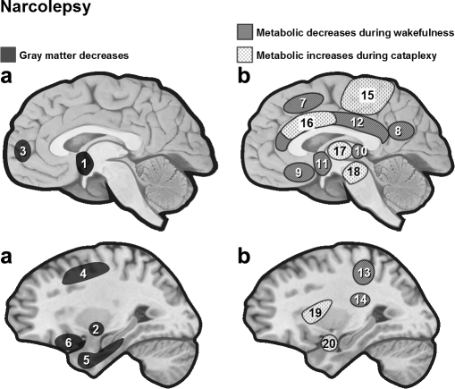
Figure 3.
(a) Anatomical brain changes in narcoleptic patients assessed by VBM. In narcoleptic patients, cortical gray matter loss was found to affect frontal brain regions,37 temporal regions,38 as well as hypothalamus, cerebellum (vermis) and ventral striatum39 (see also40). However, hypothalamus damage is not systematically found in VBM studies.36 (b) Functional brain changes in narcolepsy. Narcoleptic patients show reduced baseline activity during wakefulness (18FDG PET, SPECT) in several regions including the hypothalamus and mediodorsal thalamic nuclei.45,46 During cataplexy, one SPECT study reported hyperperfusion in several brain regions including limbic area, brainstem and motor regions51 (but see also52). 1 = hypothalamus, 2 = nucleus accumbens (right), 3 = frontomesial, 4 = prefrontal (right), 5 = inferior temporal, 6 = inferior frontal, 7 = superior frontal, 8 = inferior parietal lobule, 9 = rectal/subcallosal gyrus, 10 = dorsal thalamus, 11 = hypothalamus, 12 = cingulate, 13 = post central/supramarginal, 14 = caudate, 15 = premotor and motor cortex, 16 = cingulate, 17 = thalamus, 18 = brainstem, 19 = insula (right), 20 = amygdala (right).

