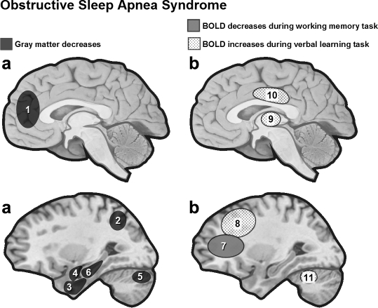
Figure 4.
(a) Regional gray matter loss in OSAS patients. VBM results in OSAS patients revealed gray matter loss limited to the left hippocampus80 or extending to regions involved in cognitive functions and motor regulation of the upper airway.79 (b) Task-related activation in OSAS patients. Functional MRI in OSAS patients during a 2-back working memory task was associated with reduced dorsolateral prefrontal activity,87 while verbal learning was associated with increases in frontal cortex, thalamus and cerebellum.88 1 = left anterior cingulate cortex, 2 = posterior lateral parietal cortex, 3 = inferior temporal gyrus, 4 = parahippocampal gyrus, 5 = right quadrangular lobule, 6 = left hippocampus, 7 = dorsolateral prefrontal cortex, 8 = inferior/middle frontal, 9 = thalamus, 10 = cingulate gyrus, 11 = cerebellum.

