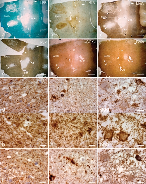
Fig. 2.
Pattern III multiple sclerosis lesions. (a–d) The brain sections containing Pattern III and Balo's type (with concentric rings of preserved myelin) active multiple sclerosis lesions are stained for LFB (a), HLA (b), MAG (c)and PLP (d). There is reduced density of LFB staining (a, asterisks) in the EA stage, whereas LFB staining is absent (a, arrows) in the LA stage of Pattern III lesions compared with NWM. The neuropathological hallmark of Pattern III lesions is the preferential loss of MAG, as indicated here by the loss of MAG immunoreactivity (c, asterisks) and intact PLP immunoreactivity (d, asterisks) in the EA stage. In contrast, MAG and PLP immunoreactivity is lost in the LA stage of Pattern III lesions (c and d, arrows). e and f: In deeper sections from the same block, there is a decrease in complex IV subunit-I (COX-I) immunoreactivity, which is diffuse in the EA stage (e, asterisks) and most prominent in the LA stage (e, arrows) compared with NWM. The porin immunoreactivity in the EA stage (f, asterisks) of Pattern III lesions is similar to the NWM. However, there is a decrease in porin immunoreactivity in the LA stage (f, arrows). Interestingly, COX-I immunoreactivity is preserved in the concentric rings of the Balo's type multiple sclerosis lesion (e). g–i: Higher magnification images show the punctate COX-I (g), porin (h) and COX-IV (i) immunoreactive elements, typical of mitochondrial staining, in the NWM (left column), EA stage (middle column) and LA stage (right column). There is a global reduction in the number of COX-I immunoreactive elements in the EA (gg) and LA (ggg) stages of Pattern III lesions compared with NWM (g). There are ramified cells consistent with microglia containing dense COX-I immunoreactivity in Pattern III lesions (gg–ggg). The number of porin immunoreactive elements appears reduced in LA (hhh), where there is tissue vacuolation, but not EA (hh) stage compared with NWM (h). The cells with enlarged cytoplasm (consistent with hypertrophied astrocytes) contain a peripheral rim of dense porin (hhh) but not COX-I (ggg) immunoreactivity, and clearly illustrate the disproportionate loss of COX-I compared with porin immunoreactivity in Pattern III lesions. The number of COX-IV immunoreactive elements is also decreased in Pattern III lesions (ii and iii) compared with NWM (i). Scale bars = 8 mm (a–f) and 10 µm (g–i).

