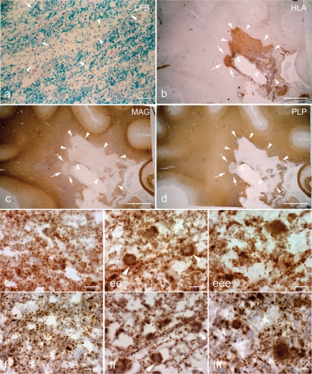
Fig. 4.
Pattern II multiple sclerosis lesions. a–d: Sections containing a Pattern II multiple sclerosis lesion are stained for LFB (a), HLA (b), MAG (c) and PLP (d). The EA stage of Pattern II lesions is identified by the perivenous demyelination, which are separated by partly preserved myelin (a, arrows), and abundance of phagocytic macrophages (b) containing myelin debris (a, arrowheads). Both MAG (c) and PLP (d) immunoreactivity are equally lost in the EA and LA stages (c and d, arrows and arrowheads, respectively) of Pattern II lesions. e and f: At higher magnification the Pattern II tissue appears vacuolated with a general reduction in the number of porin immunoreactive elements in EA (ee) and LA (eee) stages compared with NWM (e). f: The reduction in COX-I immunoreactive elements in EA (ff) and LA (fff) stage of Pattern II lesions is relative to the reduction of porin (e) elements. Scale bars = 40 µm (a), 8 mm (b–d) and 10 µm (e and f).

