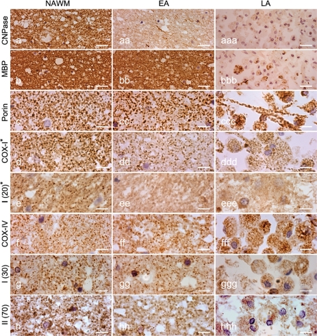
Fig. 5.
WMS lesions. The three columns show immunoreactivity in NWM (left), EA (middle) and LA (right) stages of WMS tissue. a and b: Both CNPase and MBP immunoreactivity are detectable in the NWM, whereas CNPase immunoreactivity is lost while MBP immunoreactivity is intact in the EA stage of WMS lesions. In the LA stage, both CNPase and MBP immunoreactivity are lost except within phagocytic macrophages. c: The porin immunoreactive punctate elements are numerous in the NWM and EA stage. In the LA stage of WMS lesions, there is severe tissue loss and mitochondria are mostly located in phagocytic macrophages. d–h: The number of immunoreactive elements for all mitochondrial respiratory chain complex subunits (COX-I, complex I 20 kDa or ND6, complex I 30 kDa and complex II 70 kDa) except COX-IV appears decreased in EA stage compared with NWM. Furthermore, the majority of phagocytic macrophages in LA stage show a decrease in immunoreactivity for the mitochondrial respiratory chain complex subunits (except for COX-IV) despite the abundance of porin immunoreactive elements (ccc). Asterisk indicates subunits encoded by mtDNA. Scale bars = 10 µm (a–h).

