
Figure 3.
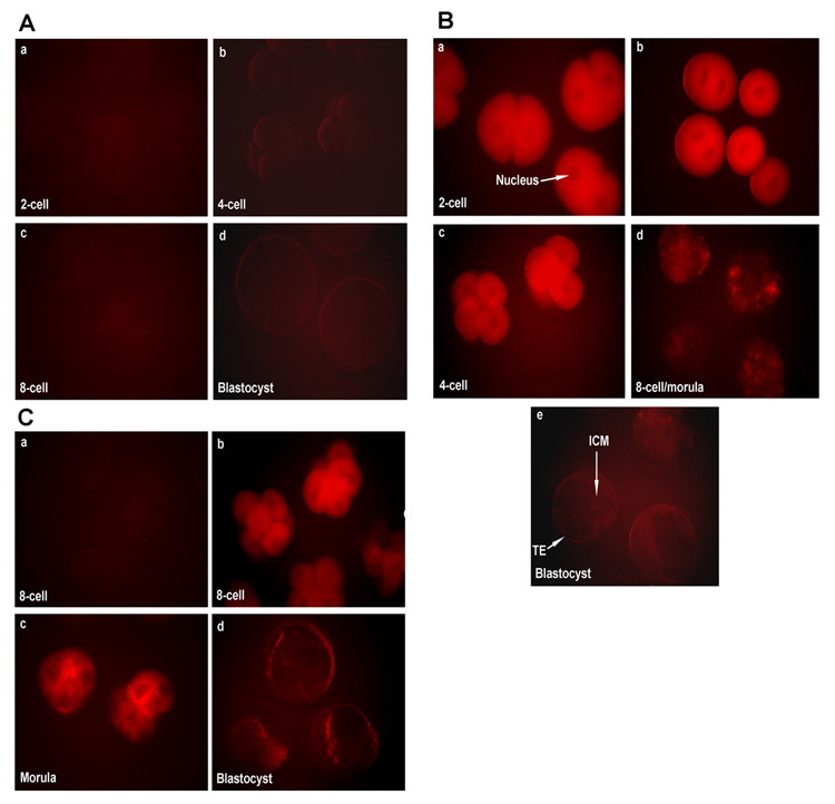
A) The level of autofluorescence in various stages of embryonic development when 2-cell embryos were only electroporated and cultured for 72 h. Autofluorescence was detected using the filter that was used to visualize Cy3-or TRITC-color. The embryonic stages (40X) are: 2-cell (a), 4-cell (b), 8-cell/morula (c), and blastocyst (d). B) Cy3 fluorescence level in various stages of embryonic development when 2-cell embryos were electroporated with Cy3-labeled control siRNA and cultured for 72 h. The embryonic stages (40X) are: 2-cell (a), fused 2-cell (b), 4-cell (c), 8-cell/morula (d), and blastocyst (e). C) Cy3 fluorescence level in the 8-cell, morula and blastocyst stages when 8-cell embryos were electroporated with Cy3-labeled control siRNA and cultured for 48 h. a, autofluorescence level in 8-cell embryos prior to electroporation; b, Cy3 fluorescence level in 8-cell stage embryos after electroporation with Cy3-labeled control siRNA; c and d, Cy-3 fluorescence level in in vitro grown morula and blastocyst stages, respectively, following electroporation of 8-cell stage embryos with Cy3-labeled control siRNA. TE, Trophectoderm; ICM, Inner Cell Mass.