
Figure 3. aMSH projections to the PVH in adult DIO and DR rats.
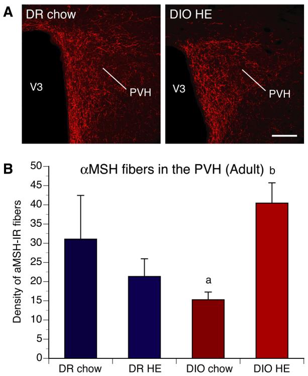
A) Confocal images and B) quantitative comparisons of aMSH-IR fibers innervating the paraventricular nucleus of the hypothalamus (PVH) of adult DIO and DR offspring of rats whose dams were fed chow or high energy (HE) diet during pregnancy and lactation. No differences in aMSH-IR fibers density in the PVH were found between DIO and DR rats overall. However, offspring of chow-fed DIO animals showed a significant reduction in the density of aMSH-IR innervating the PVH, relative to that of HE diet DIO offspring. V3, third ventricle. Scale bar, 160 um. Data are presented as mean ± SEM. P < 0.05 between a and b.