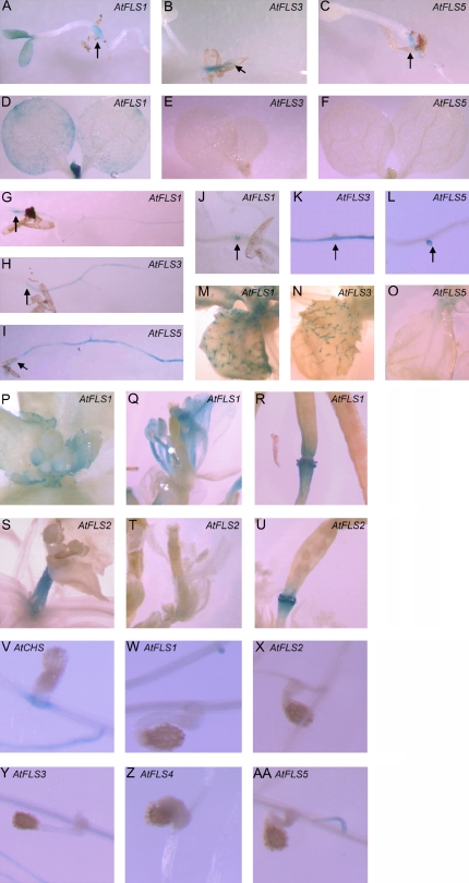
Figure 4.
Developmental, organ-specific, and parasite-induced expression of the AtFLS genes. Promoter-GUS fusions were analyzed in multiple independent transgenic lines by histochemical staining with 5-bromo-4-chloro-3-indolyl-β-d-glucuronide. Staining was observed primarily in 3-d-old seedlings (A–C), 9-d-old seedlings (D–I), initiating lateral roots in plants of various ages (J–L), trichomes on 30-d-old plants (M–O), and reproductive structures of 51-d-old plants (P–U). Arrows identify the root-shoot transition zone in A to C and G to I and lateral roots in J to L. Unlike the AtCHS promoter (V), expression of the AtFLS1 to -5 promoters was not induced by infection with the plant parasite O. aegyptiaca (W–AA).

