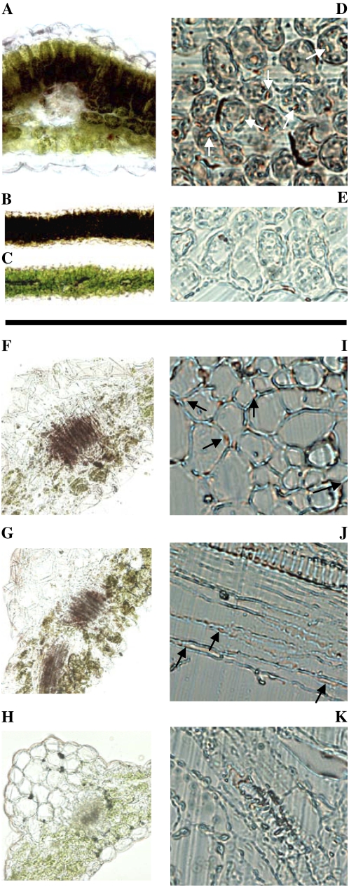
Figure 9.
Localization of β-glucosidase activity in leaves of L. japonicus wild type (A–E) and transgenic Arabidopsis expressing LjBGD2 and LjBGD4 (F–K). β-Glucosidase activity is represented by red/brown staining resulting from the hydrolysis of the artificial substrate BNG and subsequent aglucone complex formation with Fast Blue BB salt. Cross sections (80 μm) of apical leaves (fresh tissue) of L. japonicus wild type show strong color development in leaf palisade tissue in the presence of BNG and Fast Blue BB salt (A and B), while no staining is observed in the absence of BNG (C). A represents a 20× magnification, while B and C show 5× magnifications. Visualization of the subcellular localization of β-glucosidase activity is possible at 40× magnification of 6-μm cross sections of fixed apical leaf tissue. Application of BNG results in strong staining (indicated by white arrows) localized to the symplast (D), whereas only diffuse and weak apoplastic background color development is observed in the absence of BNG (E). BNG staining for β-glucosidase activity in 80-μm cross sections (20× magnification, fresh tissue) of rosette leaves of Arabidopsis expressing LjBGD2 (F), LjBGD4 (G), and the wild type (H) show that LjBGD2 and LjBGD4 both hydrolyze BNG but no BNG hydrolysis is observed in Arabidopsis wild type. I, J, and K are 40× magnifications of 10-μm sections of fixed Arabidopsis rosette leaves. I shows the subcellular localization of LjBGD2 in a cross section (black arrows). J illustrates the subcellular localization of LjBGD4 in a longitudinal section (black arrows). Very weak apoplastic background staining is observed around the xylem in Arabidopsis wild type (K) as well as in transgenic Arabidopsis. Identical subcellular and tissue localizations of LjBGD2 and LjBGD4 activity are observed upon heterologous expression of the enzymes in Arabidopsis, with activity localized in the symplast at the subcellular level (in agreement with L. japonicus) and in and around the vascular tissue at the tissue level (in contrast to L. japonicus).

