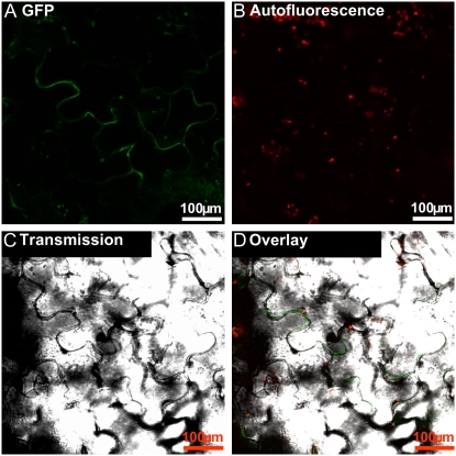
Figure 6.
Localization of a temporally expressed StAPY3-GFP fusion protein. Arabidopsis leaf epidermal cells were bombarded with a plasmid coding for the StAPY3 C-terminally fused to GFP. A, Fluorescence of GFP recorded between 500 and 525 nm. B, Autofluorescence of the chloroplasts recorded between 630 and 690 nm. C, Bright-field image of the section. D, An overlay of all three images (given in A–C). Typical examples are shown.

