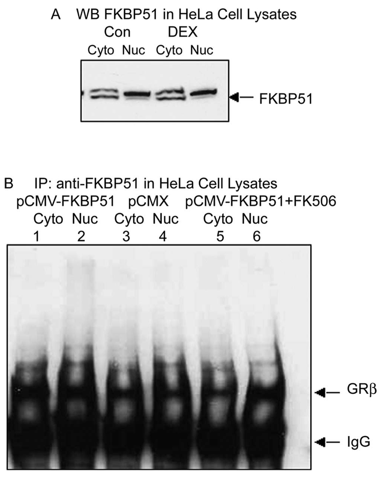
FIGURE 6.
Subcellular distribution of FKBP51 and effects of FK506 on FKBP51-chaperoned nuclear translocation of GRβ HeLa cells. (A) Confluent HeLa cells were treated with vehicle (control) or 100 nM DEX treatment for 30 minutes. The cytosol (100 µg) and nuclear (50 µg) fraction proteins were prepared and resolved on 4% to 15% SDS-polyacrylamide gradient gels. Western immunoblot analysis was performed to detect the subcellular distribution of FKBP51. (B) Subconfluent HeLa cells were transfected with either control (pCMX) or pCMX-FKBP51 plasmids and then treated with control or 1 µM FK506 for 1 hour before additional vehicle control or 100 nM DEX treatment, as labeled, for another 30 minutes. The cytosol (100 µg) and nuclear (50 µg) proteins were coimmunoprecipitated with anti-FKBP51 antibody followed by Western immunoblot analysis, to detect the effects of overexpression of FKBP51 or FK506 on subcellular distribution of GRβ. The data are representative of results of three independent experiments.

