
Fig 3. Embryoid body (EB) formation and differentiation from the parental (G-GFP) and two independent PIG-A/GPI-AP deficient hES cell clones.
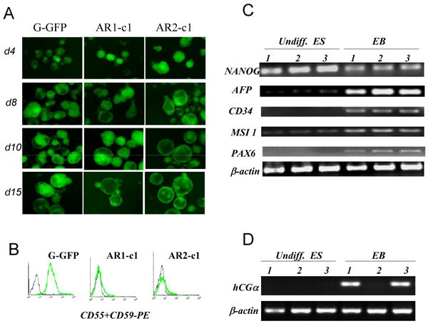
(A). Three ES cell types were cultured in suspension as aggregates to form EBs. The morphology and numbers of EBs were monitored daily for 15 days. The EB morphology is better illustrated by constitutively-expressed GFP signal. The rate as well as morphology of EBs by 3 hES cell types was similar. (B). Confirmation of the GPI-AP deficiency in EB-derived cells. Antibodies recognizing CD55 and CD59, were mixed and used to stain single cell suspension. (C). RT-PCR analysis of marker gene expression before and after EB formation at day 10. As expected, Nanog expression in G-GFP control cells (samples 1) is high in undifferentiated ES cells (undiff. ES) and visibly reduced after spontaneous differentiation by EB formation (EB). The gene expression of differentiated markers such as AFP (endoderm), CD34 (mesoderm), MSI 1 and PAX6 (ectoderm) were significantly elevated as compared to undifferentiated ES cells. AR1-c1 (samples 2) showed similar pattern to the G-GFP control, so did the AR1-c1 cells expressing PIG-A transgene (samples 3). (D). RT-PCR analysis of a marker gene expression for trophectoderm. hCGα expression from AR1-c1 hES cells (samples 2) failed to elevate after EB formation, but restored by PIG-A transgene expression. See more data by real-time quantitative RT-PCR in Supplement Fig S3.