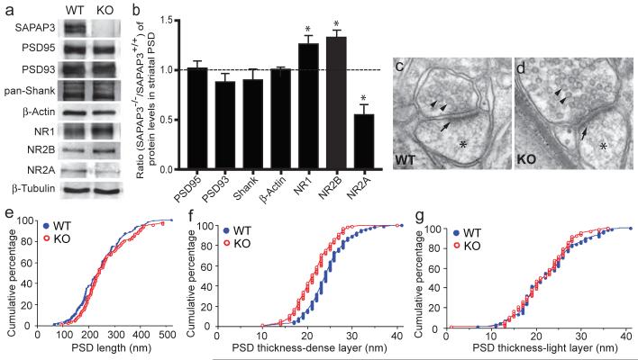
Figure 4. Structural and biochemical analyses of cortico-striatal synapses in SAPAP3 mutant mice.
a, b, The levels of PSD95, PSD93 and Shank in striatal PSD fractions are not affected in SAPAP3-/- mice. The levels of NR1 and NR2B subunits are increased, whereas that of NR2A is decreased. β-actin and β-tubulin were used as loading controls. *p < 0.05, two-tailed t test. c, d, Electron micrographs show the presence of synaptic vesicles (arrowheads), postsynaptic densities (arrows) and dendritic spines (stars). e, The length of the PSD is not significantly different in wildtype and SAPAP3-/- mice. f, g, The thickness of the dense layer (f) of the PSD in SAPAP3-/- mice, but not the light layer (g), is reduced. p< 0.001, two-tailed t test; n=94 for wildtype and n=92 for SAPAP3-/- mice.

