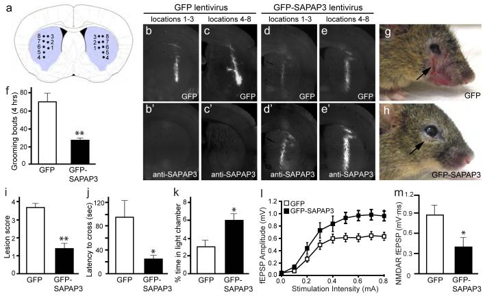
Figure 5. Lentiviral-mediated rescue of behavioral and synaptic defects in SAPAP3 mutant mice.
a, Diagram showing the approximate locations of microinjections in the striatum of SAPAP3-/- mice. Injection site 1, locations 1-3 are more anterior than injection site 2, locations 4-8. b-c’, Brain sections from a SAPAP3-/- mouse injected with GFP lentivirus show GFP fluorescence (b, c) and an absence of SAPAP3 staining (b’, c’). d-e’, Brain sections from a SAPAP3-/- mouse injected with GFP-SAPAP3 lentivirus show both GFP fluorescence (d, e) and SAPAP3 immunostaining (d’, e’). f, Compared to SAPAP3-/- mice injected with GFP lentivirus, SAPAP3-/- mice injected with GFP-SAPAP3 lentivirus showed significantly reduced over-grooming behavior. **p < 0.01, two-tailed t-test; n=8 mice/group for f, i-k. g-h, SAPAP3-/- mice injected with GFP-SAPAP3 lentivirus (h) had reduced severity of facial lesions when compared to SAPAP3-/- mice injected with GFP lentivirus (g). i, Semi-quantitative lesion scores. **p < 0.01, Mann-Whitney U test. j-k, Reduced anxiety-like behaviors in SAPAP3-/- mice injected with GFP-SAPAP3 lentivirus in the dark-light emergence test, including decreased latency to cross from the dark to light chamber (j) and increased time in the light chamber (k). *p < 0.05, two-tailed t-test. l, m, Field recordings from infected striatal area of P21-P25 SAPAP3-/- mice injected with GFP-SAPAP3 lentivirus showed an increase in cortico-striatal fEPSP amplitude (l) and a reduction of NMDAR-dependent fEPSP area (m). p < 0.001, repeated measures ANOVA for l; *p < 0.05, two-tailed t-test for m; n is 12 and 10 for l, and 10 and 9 for m for GFP injected and GFP-SAPAP3 injected, respectively .

