Figure 3.
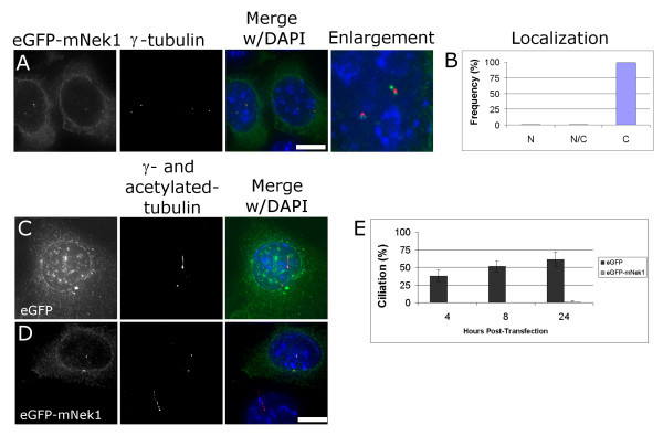
Transient overexpression of mNek1 disrupts ciliogenesis. (A) eGFP-mNek1 (green) localizes to several spots adjacent to the γ-tubulin foci (red) and is excluded from the nucleus (DAPI, blue). (B) Nuclear localization was scored as either predominantly nuclear ("N"), approximately equivalently distributed between nucleus and cytoplasm and nucleus ("N/C"), or predominantly cytoplasmic ("C"), n = 300. (C) eGFP (green) is not found associated with centrosomes and is observed in both cytoplasm and nucleus. These cells were fixed and stained 12 hours after transfection. Note the presence of a full-length primary cilium. (D) Representative fluorescent image of eGFP-mNek1 transfected cell lacking a primary cilium 12 hours after transfection. Note the ciliated wild-type cell in the same image. (E) Quantification of cells expressing a primary cilium at 4, 8 and 24 h post transfection. Error bars represent the standard deviation between two experiments, n = 200 for each experiment. Bar, 10 μm.

