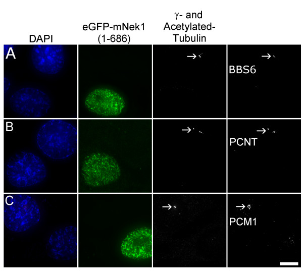
Figure 4.
Pericentriolar disruption resulting from overexpression of mNek1 (1–686) is not limited to γ-tubulin. (A-C) Representative fluorescence microscopy images of IMCD3 cells expressing eGFP-mNek1 (1–686), co-stained with acetylated- and γ-tubulin and BBS6 (A), Pericentrin (PCNT; B) or PCM1 (C). Transfected cells lack foci of these centrosomal markers. The location of the intact centrosome in the non-transfected cell is marked by an arrow. Bar, 10 μm.

