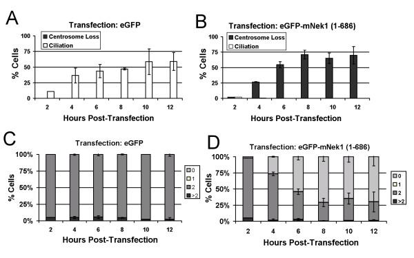
Figure 5.
mNek1 disruption of centrosomes is not due to sequential loss. Cells were scored for the presence or absence of cilia and centrosomes following transfection with eGFP-alone (A) and eGFP-mNek1 (1–686) (B). Error bars represent standard deviation of 3 experiments, n = 100 for each experiment. (C and D) Time course of γ-tubulin foci number (0, 1, 2 or > 2) in cells expressing eGFP alone (C) or eGFP-mNek1 (1–686) (D). Cells transfected with eGFP-mNek1 (1–686) are not observed to retain a single γ-tubulin focus, suggesting that loss is coincident.

