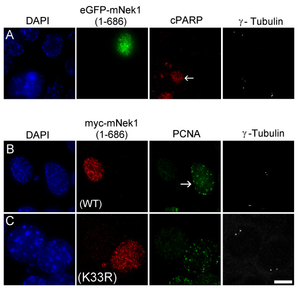
Figure 7.
mNek1-mediated centrosomal loss disrupts the cell cycle and is not a consequence of apoptosis. (A) Representative fluorescence microscopy image of IMCD3 cells transfected with eGFP-mNek1 (1–686; green), co-visualized with the apoptosis marker cleaved-PARP (red), γ-tubulin (greyscale) and DAPI (blue). Adjacent to an untransfected, representative apoptotic cell (arrow), the transfected cell lacks γ-tubulin foci but remains negative for cleaved-PARP. (B and C) Representative fluorescence microscopy images of IMCD3 cells transfected with either (B) myc-mNek1 (1–686) or (C) myc-mNek1 (1–686; K33R). α-myc (red) is co-visualized with the S phase marker PCNA (green), γ-tubulin (greyscale) and DAPI (blue). An untransfected, representative S phase cell is indicated with an arrow. 24 hours post-transfection, cells transfected with myc-mNek1 (1–686) that lack γ-tubulin foci are not observed in S phase, whereas 10% of cells transfected with myc-mNek1 (1–686; K33R) are positive for PCNA.

