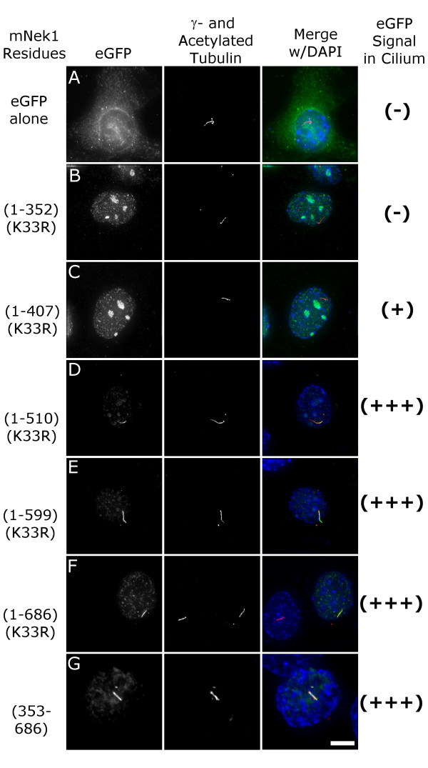
Figure 8.
The Coiled-coil domain of mNek1 contains a ciliary localization signal. (A-G) Representative images of eGFP-tagged mNek1 constructs containing the mutation K33R (green), γ- and acetylated-tubulin (red), and a merge with DAPI (blue). Localization to the primary cilium was scored by the co-localization of the acetylated-tubulin-stained cilium and the eGFP signal as either none (-), minimal (+), or strong (+++). Constructs that contain coiled-coil 2 localize to the primary cilium (D-G), and the coiled-coil domain is sufficient for localization to the primary cilium (G). Cells in these images were fixed and stained 12 hours after transfection. Bar, 10 μm.

