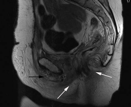
Figure 1.

T1-weighted MRI of the pelvis (sagittal view) exhibiting gross oedema of the urethra, vagina, and rectum (white arrows) with an air-fluid level within pubic symphsis extending into peritoneal retropubic space superior to the urethra (black arrow).
