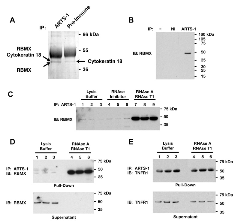
Figure 1. Co-immunoprecipitation of Endogenous RBMX and ARTS-1.

Panel A. NCI-H292 cell membrane proteins were immunoprecipitated (IP) with anti-ARTS-1 antibodies or pre-immune serum and stained with Coomassie blue. This gel is representative of two experiments. Panel B. HUVEC lysates were immunoprecipitated with the anti-ARTS-1 antibody or non-immune serum (NI) and RBMX was detected by Western blotting. Panel C. HUVEC lysates, in triplicate, were incubated with RNase inhibitor or a mixture of RNases A and T1 for 1-h prior to immunoprecipitation with the anti-ARTS-1 antibody. RBMX was detected by Western blotting. Panel D. Immunoprecipitation of proteins in HUVEC lysates were performed, in triplicate, as in Panel C. Proteins pulled-down by the ARTS-1 immunoprecipitation are shown in the top panel, while proteins remaining in lysates are shown in the bottom panel labeled supernatant. Panel E. Immunoprecipitation of proteins in HUVEC lysates, performed in triplicate, as described in Panels C and D. TNFR1 was detected by immunoblotting.
