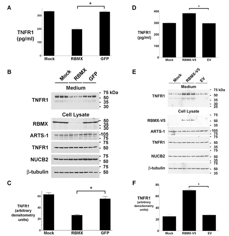
Figure 2. RBMX Modulates the Constitutive Release of TNFR1 Exosome-like Vesicles.

Panels A and B. TNFR1 in medium from HUVEC transfected with siRNA targeting RBMX or GFP was quantified by ELISA (Panel A) or Western blotting (shown in triplicate in Panel B). Mock denotes cells treated with transfection reagent alone. The asterisk denotes decreased TNFR1 release from cells transfected with siRNA targeting RBMX (P < 0.0001, n = 6). Western blots of HUVEC lysates are also shown. Panel C. Quantification of Western blots from Panel B. The asterisk indicates a significant decrease in the quantity of the 55-kDa TNFR1 present in conditioned medium from cells transfected with siRNA targeting RBMX (P < 0.003, n = 3). Panel D. The quantity of TNFR1 in conditioned medium from HUVEC transfected with plasmids encoding a V5-tagged RBMX (RBMX-V5), empty vector (EV), or the transfection reagent alone (Mock) was detected by ELISA. The asterisk denotes a significant increase in the quantity of TNFR1 present in medium from cells expressing RBMX-V5 (P < 10−9, n = 6). Panel E. Western blot showing, in triplicate, TNFR1 in HUVEC conditioned medium (top panel) and RBMX-V5 (detected with an anti-V5 antibody), ARTS-1, TNFR1, NUCB2, and β-tubulin in cell lysates (bottom panel). Panel F. Quantification of Western blots from Panel E by densitometry. The asterisk denotes significant increase in the quantity of 55-kDa TNFR1 present in medium from cells transfected with the RBMX-V5 plasmid (P < 10−4, n = 3).
