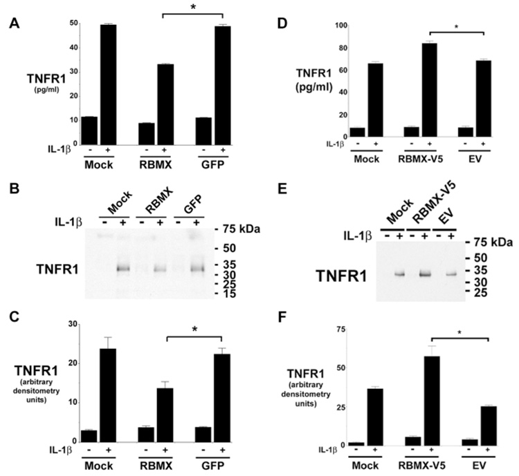
Figure 3. Down-regulation of RBMX Expression by RNA Interference Attenuates the IL-1β-mediated Proteolytic Cleavage of TNFR1 Ectodomains.

Panels A and B. HUVEC transfected with siRNA targeting RBMX or GFP were treated with 20 ng/ml of IL-1β for 2-h and the concentration of TNFR1 in conditioned medium was determined by ELISA (Panel A) or Western blotting (Panel B). Mock denotes treatment with transfection reagent alone. The asterisk indicates that the quantity of TNFR1 in conditioned medium after IL-1β stimulation from cells transfected with siRNA targeting RBMX was significantly reduced (P < 0.0001, n = 6). The blot in Panel B is representative of three individual experiments. Panel C. Quantification of Western blots from Panel B by densitometry. The asterisk indicates a significant decrease in the quantity of the 34-kDa sTNFR1 in conditioned medium after IL-1β stimulation from cells transfected with siRNA targeting RBMX (P < 0.02, n = 3). Panels D – E. HUVEC were transfected with plasmids encoding V5-tagged RBMX (RBMX-V5), the empty vector (EV), or the transfection reagent alone (Mock) for 2 days prior to the addition of fresh medium, without or with 20 ng of IL-1β for 2-h. The quantity of TNFR1 present in conditioned medium was detected by ELISA (Panel D) and Western blotting (Panel E). The asterisk denotes a significant increase in the quantity of TNFR1 present in medium after IL-1β stimulation from cells transfected with the RBMX-V5 plasmid (P < 0.00015, n = 6). This blot in Panel E is representative of three individual experiments. Panel F. Quantification of Western blots from Panel E by densitometry. The asterisk denotes a significant increase in the quantity of 34-kDa TNFR1 present following IL-1β stimulation from cells transfected with the RBMX-V5 plasmid (P < 0.008, n = 3).
