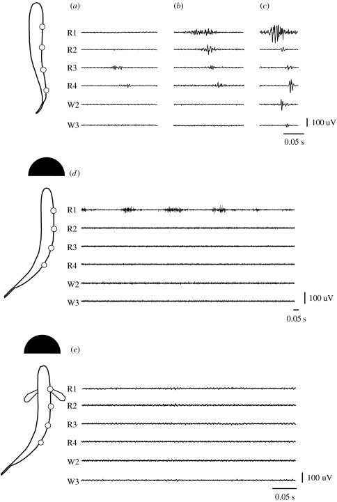
Figure 9.
Red and white axial muscle EMG traces of a trout swimming in uniform flow at: (a–c) different speeds, and in a turbulent vortex street behind a cylinder (d) without and (e) with pectoral fin activity. Red muscle electromyograms (R1–R4) are approximately aligned with the electrode positions along the body outlines to the left, with the W2 and W3 white muscle electromyograms corresponding to the same longitudinal placement as R2 and R3 insertion sites (Liao 2004). (a) When swimming in uniform flow at 1.8 L s−1, red muscle activity propagates down to the posterior end of the body. (b) At 3.5 L s−1, a wave of red muscle activity travels the entire length of the body. (c) At 5.0 L s−1, white muscle is recruited. (d) Vortex street 3.5 L s−1. The same fish Kármán gaiting behind a cylinder placed in a 3.5 L s−1 flow activates only its anterior-most red muscles (R1). Several swimming cycles are shown to illustrate the rhythmicity and variation of activity. (e) Vortex street with pectoral fin activity 3.5 L s−1. At times during the Kármán gait when the pectoral fins are active, there is no appreciable axial muscle activity along the entire body. Black scale bars are given for time (s) and muscle activity (uV).

