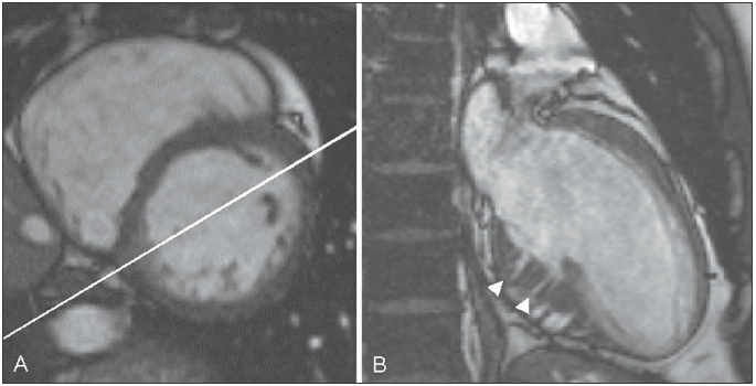
Figure 1.

End-diastolic short-axis (A) and dedicated long-axis (B) cine images of a hypertrophic cardiomyopathy mutation carrier. Note the bright triangular spot in the inferoseptum (A) through which the dedicated long-axis image is planned. The white arrowheads indicate crypts in the inferoseptum of the left ventricle (B).
