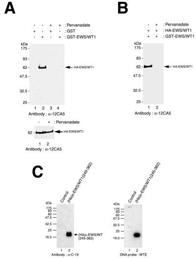
Figure 5.
Phosphorylation of EWS/WT1 inhibits self-association. (A) Loss of EWS/WT1 self-association after phosphorylation. Cell lysate containing HA-tagged EWS/WT1 was incubated with either GST-EWS/WT1 or GST alone (indicated above panel). After affinity-selection, HA-tagged EWS/WT1 protein was quantitated by Western blot analysis using the anti-HA antibody, 12CA5. Treatment of cells with vehicle or with pervanadate is indicated at the top. The arrow indicates the position of migration of HA-EWS/WT1. Molecular mass markers are provided to the left and are derived from prestained NEB protein standards (broad range). (Lower) Western blotting shows that similar amounts of HA-EWS/WT1 are present in the input lysates. Ten percent of the input samples were separated on a 10% SDS/PAGE, transferred onto Immobilon PVDF membrane, and probed with anti-HA antibody, 12CA5. (B) EWS/WT1 self-association is not increased after phosphorylation. Ten micrograms of pcDNA3:HA-EWS/WT1 and pcDNA3:GST-EWS/WT1 was cotransfected into COS-7 cells and half of the cells were treated with pervanadate for 15 min before harvesting. After selection with glutathione-Sepharose beads, proteins were separated by 10% SDS/PAGE and interrogated by Western blotting using an anti-HA antibody (12CA5). Whether cells were pretreated with vehicle (−) or pervanadate for 15 min (+) before extracts were prepared is indicated at the top. (C) EWS/WT1 self-association and DNA binding domains are separable. A deletion mutant of EWS/WT1 containing six histidines, (His)6-EWS/WT1(245–362), was expressed in Escherichia coli, purified with Ni-nitrilotriacetic acid agarose resin, and probed with α-C-19 (Left). After transfer to nitrocellulose, Southwestern blot analysis was performed with WTE as probe (Right). The position of molecular mass markers are indicated on the left (NEB broad range markers) and the position of migration of (His)6-EWS/WT1(245–362) protein is indicated on the right.

