
Figure 6.
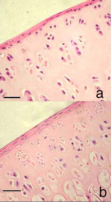
Histological sections of cartilage obtained from the distal femur of a rat at 23 weeks of age. 4a. This cartilage has been not been impacted i.e. is a control section but has been cultured for 48 hours. Few chondrocyte clusters (defined as cell groupings of 4+ nuclei, not arranged perpendicular to the joint surface) are present. 4b This cartilage has been subjected to single impact load of 200 g from 8 cm and cultured for 48 hours. There are significantly more chondrocyte clusters in this section than in the control section. Both sections are stained with H&E. Scale bar = 15 μm.